Stapeliad Flowers
|
|
|
|
published in 11. Info-Brief of the IG Ascleps S. 5-9 (2003), updated
Friederike Hübner and Ulrich Tränkle
|
|||||||||||||||||||||||||||||||||
|
|
|
|
After publication of the last circular letter I received an email from a fellow member. He had read the articles with interest and said he loves sowing, but was not by any means able to assign the names from the text to the parts of the flowers without ambiguity. I had to admit that he was right. Without pictures the flower structure of asclepediceae is difficult to understand. I would therefore like to explain it in more detail. Yet, when it comes to terminology, different authors use various terms when describing a flower structure.
Tab.1 (on the left) gives an overview of established terms without claiming completeness |
|||||||||||||||||||||||||||||||||
|
|
Ill. 1: General flower structure of genus Huernia exemplified through Huernia boleana |
|
Illustration 1 is a longitudinal section of Huernia boleana. The cream coloured flower has quite long papillae, which bear dark red dots or stripes, at least at the end. The staminal column sits at the lower end in the centre of the corolla. The outer corona lobes form a basal dark disk. The inner corona lobes are prolonged, leaning towards each other over the stigma head and then diverging again. Tiny tubercles make their upper ends coarse. |
|||||||||||||||||||||||||||||||||
|
|
Ill.
2: Detail of
Ill.1. |
|
Illustration 2 shows the staminal column of Huernia boleana. The reproductive organs are located between the inner corona lobes. The guiding rail bears a wide funnel-shaped opening at the lower end, which makes the threading of the pollinii easier. The stigma is hidden and sits inside at the upper end of this guiding rail. This is where the pollinarii are located as well: The pollinii are often protected by membranes, of which two each are connected by the translator, which is also connected to the staminal column at the upper end of the guiding rail.
|
|||||||||||||||||||||||||||||||||
|
|
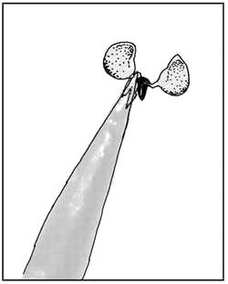
Ill. 3: Pollinarium fixed with glue |
|
Illustration 3 shows a pollinarium glued to the tip of an insect needle in order to facilitate hand pollination. |
|||||||||||||||||||||||||||||||||
|
|
Ill. 4: Flower structure of genus Larryleachia exemplified through Larryleachia cactiformis. |
|
Illustration 4 illustrates the flower and bud of Larryleachia cactiformis. The crown is patterned rich in contrast, it is tuberculate and displays a tureen-shaped hollow in the centre. The staminal column shows remarkably cornuted outer corona lobes. The inner corona lobes meet at the stigma head or overlap slightly, yet they do not rise upwards. |
|||||||||||||||||||||||||||||||||
|
|
Ill. 5: Threaded pollinium in Larryleachia picta. |
|
Illustration 5 is highly magnified and shows a threaded pollinium in Larryleachia picta. You can clearly see the oblique angle, which must be kept up when threading the guiding rail. The lower and the left pollinarii are removed. |
|||||||||||||||||||||||||||||||||
|
|
Ill. 6: Flower structure of Larryleachia marlothii. |
|
Illustration 6 shows the flower structure of Larryleachia marlothii in comparison to illustration 4. The horns of the outer corona lobes are crescent-shaped, yet instead of a central horn they only have slight bumps. The pollinarii and the guiding rail are also easily accessible. The inner corona lobes of Larryleachia marlothii are long, rise over the stigma head and meet at the tips. The crown is bowl-shaped as well until halfway up the corolla lobes. The surface is as coarse as sand paper.
|