published in Info Letter N° 9, IG Ascleps, p. 14-15 (2002)
by Friederike Hübner
During the early years of my studies in biology, I focused on plant anatomy to a great extent. Microscopes open up a world of its own, mirroring adaptation processes and environmental conditions condensed in the cellular structure of a plant in its habitat. Thus I started to survey the genera Ceropegia and Cynanchium in more detail and concentrated on the morphology of grown shoots.
Thus, I would like to describe the anatomy of Ceropegia linearis ssp. woodii, which to me is an extraordinary beautiful species of Ceropegia. This probably accounts for its wide distribution as a plant for hanging baskets.
Ceropegia linearis ssp. woodii has relatively long internodes (the distance from one pair of leaves to the next) if it grows healthily. Its shoots are comparatively slender. If you have ever tried to tear apart a shoot, you will have found it is much harder than expected. This phenomenon can be explained by looking at the cellular structure of shoots.
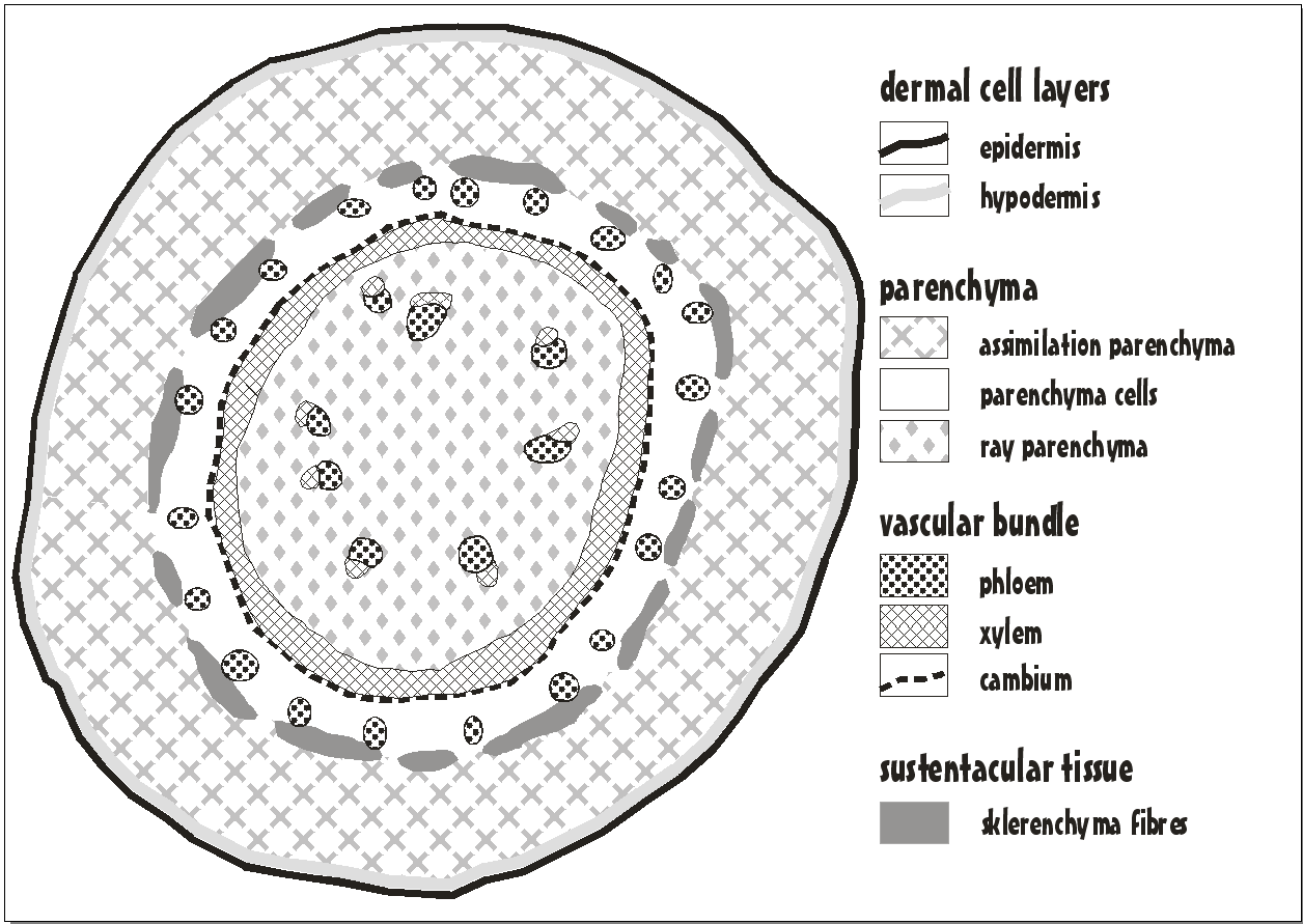
Ill. 1 Shows a semi-schematic overview of a shoot cross section.
Ill. 2 Shows a blown-up section on cellular level.
On the outside there is a cellular layer, the epidemis ("outer skin"), sealing the shoot off. There follows a second complete layer of cells called hypodermis ("inner skin"). Bother cell layers do not hold any chloroplasts (green cell bodies providing photosynthesis), but red embeddings which might be grease droplets.
Then comes a thick cellular layer made up of big cells with plenty of chloroplasts and vast intercellular spaces. Depending on the amount of solar radiation, there also appear some red central vacuoles. These are big, often coloured spaces filled with water and refuse materials in the inner region of the grown-up cell, whereas the effectively vital cytoplasm clings to the fringe.
The system of vascular strands is developed in the form of a closed ring, protecting the phloem (living cells for the transport of saccharides and other cellular components) from too much shrinkage through sklerenchyma fibres which are very hefty in cross section and extremely elongated and acicular in longitudinal section. These fibre cells are the reason for the sturdiness of the long shoots. The airless xylem transports water as well as micronutrients and macronutrients. It is rather hefty as well, since the transport is kept going almost exclusively by negative pressure. Thus, the xylem cannot collapse even if the water situation is very tight – the water flow never gets ruptured. There is a cellular layer between phloem and xylem, called the cambium, lining out new lines of cells towards the outside and the inside. This enables the plant to develop shoots that get a little bit thicker by-and-by.
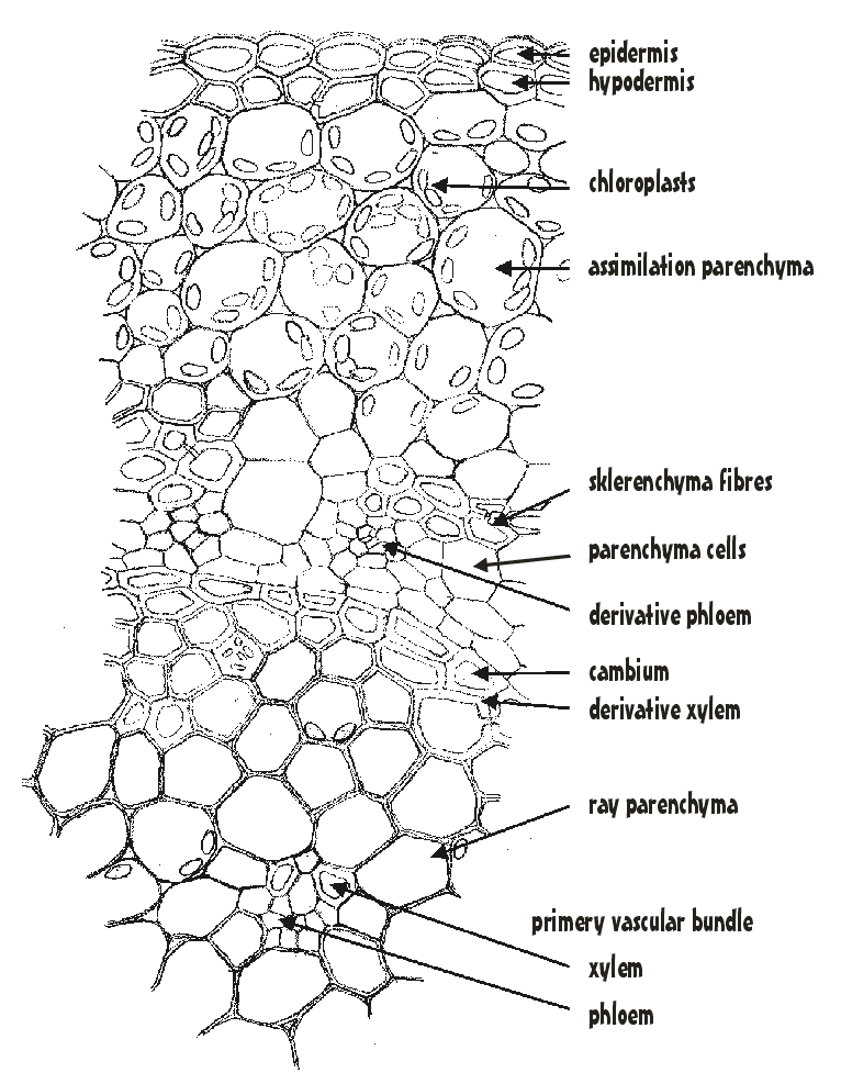
Inside the shoot there is the ray parenchyma (thin-walled cells with intercellular spaces), in which there are some singular vascular bundles left over from very early phases of growth. The plant embeds starch granules (leucoplasts) in this ray parenchyma, functioning as nutriment repository for hard times. I once cut off a shoot that had stopped growing long ago and was not able to perceive the cellular structure for all the leucoplasts that came falling out. This also accounts for the fact that Ceropegia cions stemming from old shoots can grow new rooting rather quickly – they simply have enough resources.